
Vulval & Vaginal Varicose Veins
Vaginal varicose veins can develop when pelvic or ovarian veins allow blood to flow backwards, increasing pressure in the veins around the vagina, vulva and upper legs. They often become noticeable during pregnancy, when hormonal and physical changes cause pelvic veins to dilate. After childbirth, the veins may become smaller, but they do not always disappear completely. Accurate diagnosis is important because pelvic venous reflux requires a different approach from ordinary leg varicose veins. Understanding the symptoms, causes, role of pregnancy, ovarian veins, diagnosis and treatment can help patients receive appropriate care.
What Are Vulval and Vaginal Varicose Veins?
Vulval varicose veins are enlarged veins around the vulva, while vaginal varicose veins involve veins within or around the vaginal tissues. Both can develop when blood flows backwards through the pelvic veins instead of travelling normally towards the heart.
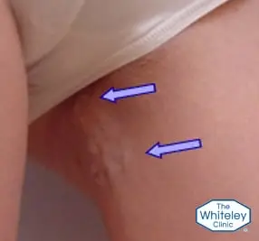
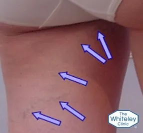
The problem often starts deeper inside the pelvis. Reflux may occur in the ovarian veins or internal iliac veins. Increased venous pressure can then extend into veins around the vagina and vulva. In some women, the abnormal veins remain mainly around the vulva. In others, they extend into the upper inner thigh, down the inner leg or towards the back of the thigh.
The distribution of the veins provides an important clinical clue. Veins appearing behind the adductor longus tendon are often associated with pelvic or ovarian vein reflux. By contrast, veins arising in front of this tendon are more likely to have a conventional leg-vein source.
Vaginal varicose veins can also be difficult to recognise because the abnormal veins may lie deep within the pelvis. Some women have no visible veins or significant symptoms until pregnancy increases pressure within the pelvic venous system.
What Symptoms Can Vulval Varicose Veins Cause?
Vulval and vaginal varicose veins can cause visible swelling, aching or discomfort around the vulva and vagina. Symptoms may become more noticeable after prolonged standing or during pregnancy.
Pelvic venous reflux can also increase pressure on nearby organs. Possible symptoms include:
- Pelvic pain or aching
- A dragging or heavy sensation in the pelvis
- Worsening stress urinary incontinence
- Worsening symptoms of irritable bowel syndrome
- Discomfort around the vulva or vagina
- Visible or swollen veins around the labia
Symptoms can also vary during the menstrual cycle. Hormonal changes affect the pelvic veins, so discomfort may become worse around the time of a period.
Not every woman with pelvic venous reflux develops symptoms. Many women remain unaware of the problem until pregnancy makes the abnormal veins more apparent. As a result, vaginal varicose veins can sometimes exist without obvious external signs.
Can Pregnancy Make Vulval and Vaginal Varicose Veins Worse?
Pregnancy has an important role in the development and worsening of vulval and vaginal varicose veins. Hormonal changes affect the vein walls, while the growing uterus increases pressure within the pelvic venous system. Consequently, pelvic veins can become considerably wider during pregnancy.
This increased pressure may open collateral veins that were previously less noticeable. Abnormal blood flow can then extend from the pelvis into veins around the vagina, vulva and legs.
After childbirth, hormone levels fall and pressure from the uterus is removed. Therefore, vaginal varicose veins and vulval varicosities often become smaller. However, a vein that has developed persistent reflux may remain abnormal because its valves no longer prevent backward blood flow.
If this reflux remains untreated, abnormal veins can continue to spread into the leg. Further pregnancies can increase pelvic venous pressure again and may worsen an existing condition.
In some women, the veins become so distended during pregnancy that they do not return to their previous size. Large, visible and occasionally painful varicosities can then remain around the vulva or vagina.
Although pregnancy is strongly associated with this condition, it is not limited to women who have been pregnant. The Whiteley Clinic has identified two women with the condition who had never been pregnant.
What Causes Varicose Veins in This Area?
The underlying problem is usually venous reflux. Healthy veins contain valves that help direct blood towards the heart. When the valves in pelvic or ovarian veins become incompetent, blood can flow backwards. This increases venous pressure and causes the veins to enlarge.
Ovarian vein reflux is an important cause, but it is not the only possible source. Reflux in the internal iliac veins can also produce abnormal pelvic venous flow. A detailed assessment is therefore needed before treatment is selected.
The position of visible leg veins can provide another important clue. Veins on the inner thigh that appear behind the adductor longus tendon, or veins extending towards the back of the thigh, may indicate reflux from pelvic or ovarian veins.
Vaginal varicose veins should not automatically be treated as ordinary leg varicose veins. Removing or injecting the visible veins without identifying their source may provide temporary improvement while leaving the underlying reflux untreated.
Understanding Ovarian Veins and Ovarian Varicocele
The ovaries are the female gonads and are comparable to the testes in men. During foetal development, the ovaries initially form high in the abdomen near the kidneys. They later descend into the pelvis, taking their venous drainage with them.
The ovarian veins can develop valve problems in a similar way to the testicular veins in men. When an ovarian vein becomes incompetent and allows blood to reflux, enlarged veins can develop around the ovary. This is sometimes described as an ovarian or pelvic varicocele.
Ovarian or pelvic varicocele can cause a dull ache or dragging sensation, particularly around menstruation. The enlarged pelvic veins can also spread within the pelvis and put pressure on the bladder or bowel.
There are two important differences in women. First, the veins can spread further through the pelvis and affect nearby organs. Secondly, these abnormal veins lie inside the body and are therefore not easily visible during a routine examination. Symptoms may consequently be overlooked or attributed to non-specific women's health problems.
How Are Vulval and Vaginal Varicose Veins Graded?
A clinical grading system helps clinicians describe the severity of vulval varicosities and communicate consistently when planning treatment. Before 2012, there was no established clinical grading scale specifically for these veins.
The Whiteley Clinic grading scale was published in early 2012 and classifies the condition into five grades:
Grade 0 – Normal
No vulval varicosities and no venous reflux are present.
Grade 1 – Venous reflux without visible vulval varicosities
No visible varicose veins are present in the vulva, but ultrasound confirms reflux in the vulval veins. This is usually associated with para-vulval varicose veins on the inner thigh. The original Whiteley Clinic data described this as common, occurring in approximately 1 in 7 women presenting with leg varicose veins and 1 in 5 women after vaginal delivery.
Grade 2 – Visible vulval varicosities
Varicose veins can be seen through the mucosa of the inner labia and lower vagina, with ultrasound-confirmed reflux in the vulval veins.
Grade 3 – Isolated vulval varicosities
Varicose veins are visible through the skin of the outer labia majora when standing, without distortion of the general anatomy.
Grade 4 – Extensive vulval varicosities
Widespread varicose veins of the labia cause distortion of the skin and overall anatomy when standing.
This scale helps distinguish hidden venous reflux from more extensive visible disease.
How Is the Diagnosis Made Accurately?
Diagnosis begins with a clinical assessment. A history of vulval varicosities during pregnancy can provide an important clue. The distribution of visible veins also helps determine whether their source is likely to be in the leg or pelvis.
When pelvic or ovarian vein reflux is suspected, a specialised ultrasound examination may be necessary. Standard abdominal ultrasound cannot always show the pelvic veins clearly because they lie deep within the pelvis.
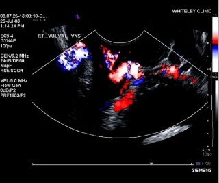
A transvaginal duplex ultrasound provides a more direct assessment. A specialist vascular scientist uses a vaginal ultrasound probe to examine the pelvic veins and assess blood flow. The examination can identify pelvic varicose veins and help determine where the reflux originates.
The scan can assess the ovarian veins as well as other pelvic veins, including the internal iliac veins. The findings can also be demonstrated to the patient during the examination. A detailed report then helps the clinical team plan appropriate treatment.
The examination becomes particularly important when varicose veins appear behind the adductor longus tendon. These veins may originate from pelvic or ovarian reflux and may not respond to conventional treatment for ordinary leg varicose veins.
Why Can Incorrect Treatment Lead to Recurrence?
Not all vulval or vaginal varicose veins have the same underlying cause. If clinicians simply remove or inject visible veins without treating the underlying pelvic reflux, the abnormal venous pressure can remain.
Historically, surgeons treated the pelvic veins directly, using both open and laparoscopic procedures. These operations could be traumatic, and the results were often less satisfactory.
Some treatments focus only on the visible veins in the upper thigh. Removing or injecting these veins can make them disappear for a time. However, the underlying problem may persist. If ovarian or pelvic reflux continues, new veins can develop, or previously treated veins may reappear.
Vaginal varicose veins can therefore return even after treatment initially appears successful. The visible vein is not always the source of the problem. For a longer-lasting result, clinicians need to identify and treat the underlying source of the reflux.
How Are Vulval and Vaginal Varicose Veins Treated at the Whiteley Clinic?
The Whiteley Clinic has studied pelvic and vulval venous disease since 1999 and has developed a protocol-based approach to diagnosis and treatment. The process combines detailed vascular assessment with minimally invasive treatment and involves a vascular surgeon, vascular scientist and interventional radiologist.
When ovarian or pelvic venous reflux requires treatment, clinicians may use coil embolisation. This minimally invasive technique uses X-ray guidance to access the affected vein. The clinician inserts a needle to gain access to the vein and then guides a thin catheter towards the vein responsible for the abnormal blood flow. Once the catheter reaches the correct position, the clinician places small metal coils inside the vein to block the abnormal reflux.
Coils are not a new technology. Clinicians have used them for many years to block blood vessels in different parts of the body. They also have a long history of use in treating testicular varicocele, and specialists have extensive experience using them to treat ovarian vein reflux.
However, the technique involves more than simply placing coils. Accurate diagnosis, appropriate patient selection and precise positioning all play an important role. After identifying the veins responsible for the abnormal blood flow, the interventional radiologist places the coils under X-ray guidance.
For patients with vaginal varicose veins, the aim is not simply to remove visible veins. Clinicians must first identify the underlying source of the abnormal venous flow so that treatment can target the reflux.
The Whiteley Clinic approach combines specialist imaging, a protocol-based assessment and targeted minimally invasive treatment. Research published by Professor Whiteley and members of the clinic has contributed to understanding this condition, with further research continuing to assess treatment outcomes.
Getting the Right Diagnosis Matters
Vulval and vaginal varicose veins can be uncomfortable, distressing and difficult to diagnose. Pregnancy often brings the condition to attention, but pelvic venous reflux can persist after childbirth and may later contribute to varicose veins in the legs.
Because pelvic venous disease can produce symptoms that overlap with other conditions, accurate assessment is important. A detailed history, physical examination and specialised transvaginal duplex ultrasound can help identify the source of reflux.
When treatment focuses only on visible veins, recurrence can occur. A source-directed approach instead looks for ovarian or pelvic venous reflux and considers appropriate minimally invasive treatment.
For patients with vaginal varicose veins, understanding where the abnormal blood flow starts is therefore a key part of effective management.